
Introduction: We’ve Been Measuring the Wrong Thing
In sports medicine, we often rely on familiar markers:
Range of motion
Perceived tightness
Performance outputs
These are useful but they’re indirect.
They tell us something changed, but not what actually changed at the tissue level.
A recent study titled “Effects of Lower-Leg Fascial Flossing on Flexibility and Performance in Collegiate Distance Runners” takes a different approach.
Instead of just asking:
“Does it feel better?”
It asks:
“Is tissue behavior actually changing?”
And more importantly:
Can we measure that with ultrasound?
The Study: Moving Beyond Subjective Measures
This was a randomized cross-over study in nationally competitive distance runners.
Each athlete:
completed a fatigue protocol (treadmill running)
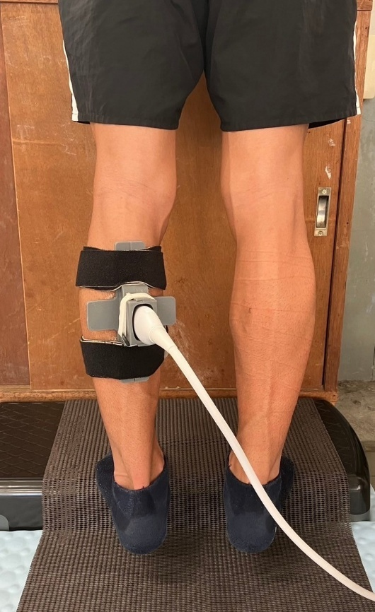
received fascial flossing on one leg
used the other leg as control
They measured:
Ankle dorsiflexion (WBLT)
Perceived tightness (VAS)
Reactive Strength Index (RSI)
Myofascial gliding via ultrasound
That last one is where things get interesting.
Key Findings: Not Just Feeling Better - Moving Better
After fatigue:
Flossed leg:
↓ perceived tightness
↑ dorsiflexion ROM
maintained jump performance
improved myofascial gliding
Control leg:
↑ perceived tightness
↓ RSI (performance drop)
no meaningful change in gliding
The critical finding:
Only the flossed limb demonstrated improved tissue gliding on ultrasound
What Is “Fascial Gliding” (Clinically Speaking)?
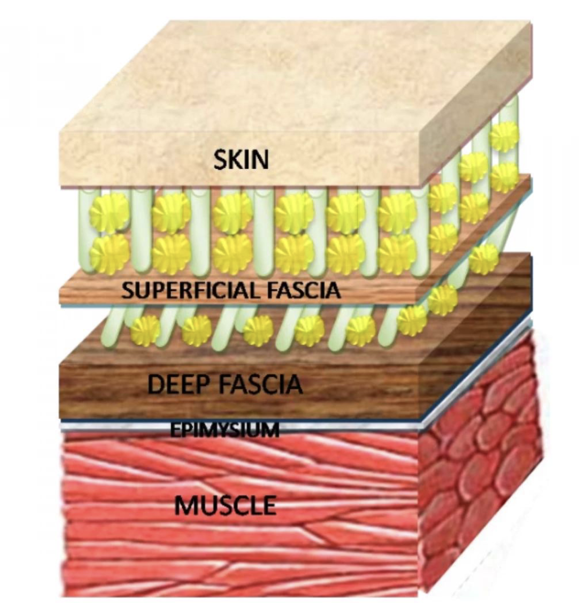
Fascia is not a static structure.
It is:
layered
continuous
and designed for relative motion between tissues
Healthy systems: muscle and fascia move independently and smoothly
Dysfunctional systems: tissues move together as a unit (reduced glide)
How Did They Measure It?
This is where the study stands out.
They used:
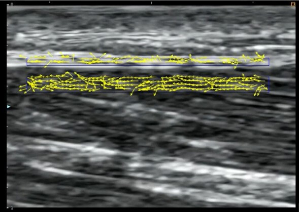
Ultrasound + optical flow analysis
Specifically:
tracked motion of superficial fascia and underlying muscle
calculated cross-correlation of movement velocity
Interpretation:
High correlation → tissues moving together → poor gliding
Low correlation → independent motion → better gliding
This is a shift from:
static imaging
to
dynamic tissue behavior analysis
Why This Matters for Clinicians
This changes how we think about common complaints like:
“tight calves”
“stiff lower leg”
“heavy legs after training”
These may not just be:
metabolic fatigue
neuromuscular changes
They may reflect: altered mechanical interaction between tissues
The Mechanism (What Might Be Happening?)
The authors suggest several contributors:
mechanical compression → altered fascial viscosity
improved tissue sliding (thixotropic effects)
changes in mechanoreceptor input
possible circulatory/reperfusion effects
The key idea:
Interventions may influence how tissues move relative to each other not just how they feel
The Bigger Shift: From Structure → Behavior
Traditionally, ultrasound has been used to assess:
thickness
echogenicity
structural abnormalities
But this study highlights a growing direction:
Ultrasound as a tool for measuring tissue behavior
Not just:
what tissue looks like
But:
how it moves
how it interacts
how it adapts under load
Where This Connects to the Future
This is where things get even more interesting.
This study used:
motion tracking (optical flow)
cross-correlation
Other emerging approaches (including what we’re working on) are exploring:
frequency-domain analysis (FFT)
structural organization metrics (PSFR-like)
quantitative tissue profiling
Different methods but same goal:
Moving from subjective interpretation → objective quantification
Clinical Takeaways
Fascial flossing may:
improve flexibility
reduce perceived tightness
help maintain performance post-fatigue
More importantly:
it may influence tissue mechanics at a deeper level
Ultrasound is evolving:
from imaging tool
to measurement tool
Limitations (Important to Acknowledge)
small sample size (n = 17)
male endurance athletes only
short-term effects only
technique-specific variables (pressure, application)
This is not a definitive answer.
It is an important step forward.
Final Thought
We are entering a new phase in musculoskeletal ultrasound:
Not just seeing tissue But measuring how it behaves
And once we can measure behavior:
we can start tracking it we can start comparing it and eventually… we can start understanding it in ways we couldn’t before
If You’re a Clinician
The question isn’t:
“Does this technique work?”
The better question is:
“What is it changing at the tissue level and can we measure it?”
Because that’s where the future is heading.
If You’re Interested in This Space
If you’re a clinician working in sports medicine or performance:
How are you currently assessing tissue changes?
Are you relying on subjective measures, or objective tools?
What would change if you could track tissue behavior over time?
We’re continuing to explore this area through:
education
clinical application
and emerging technology
If this is something you’re interested in, feel free to reach out or follow along as we continue to build in this space.