
Knee ultrasound can help answer focused clinical questions: Is swelling intra-articular or bursal? Is the extensor mechanism intact? Does an apparent tendon abnormality persist after correcting the insonation angle?
For clinicians performing musculoskeletal ultrasound, diagnostic confidence depends on accurate anatomical localization, reproducible technique, and recognition of what the examination cannot establish.
A 2026 clinical review by Sachdeva and Rao discusses knee point-of-care ultrasound, including effusions, bursitis, tendon pathology, meniscal and collateral ligament assessment, and procedural applications.[1] The following clinical interpretation emphasizes the findings and pitfalls most relevant to advanced MSK ultrasound practice.
Start with anatomy and image quality
Position the patient supine with the knee slightly flexed for the anterior examination. A small support beneath the knee can improve comfort and access to the quadriceps tendon, suprapatellar recess, and patellar tendon.
Select a transducer frequency appropriate for the target depth and optimize gain, depth, and focal position. Examine structures in both longitudinal and transverse planes, following their course rather than relying on a single static image.
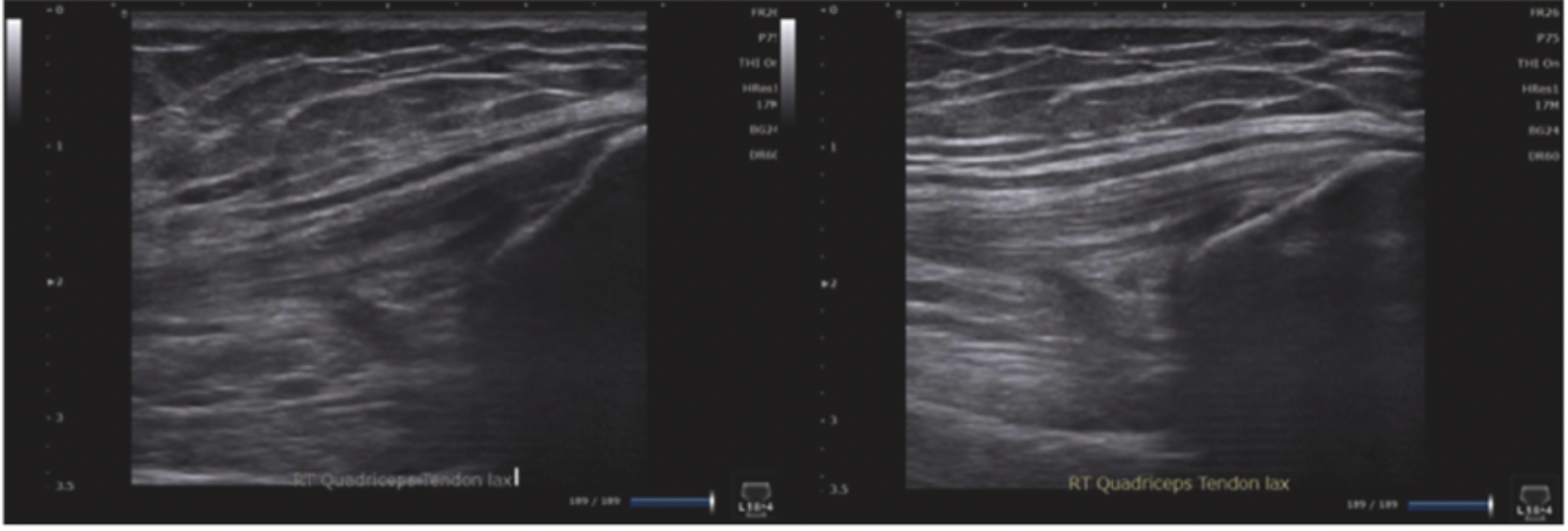
Before diagnosing tendon pathology, correct for anisotropy. Tendons can appear hypoechoic when the ultrasound beam is not perpendicular to their fibers, particularly near curved insertions. Adjusting the probe angle may restore the normal fibrillar appearance.
A hypoechoic region that disappears with angle correction should not be interpreted as a tear.
Source: Sachdeva S, Rao VV. Point-of-Care Ultrasound of the Knee: Diagnostic Applications, Procedures, and Best Practices. Journal of Radiology Nursing. 2026;48:101467. Figure 11. https://doi.org/10.1016/j.jradnu.2026.101467
Localize fluid before assigning a diagnosis
The clinical description “swollen knee” does not identify the affected compartment.
Assess the suprapatellar recess and accessible joint recesses, then determine whether additional fluid lies within a separate bursa or another periarticular structure. In the suprapatellar region, localize fluid relative to the quadriceps tendon, suprapatellar fat pad, prefemoral fat pad, and femur.
Describe the collection’s location, extent, and internal appearance. Fluid may be anechoic or contain internal echoes; echogenicity alone cannot determine its cause.
For a hot, acutely painful joint, identifying an effusion does not establish or exclude septic arthritis. Clinical assessment and, when indicated, synovial fluid analysis remain necessary.
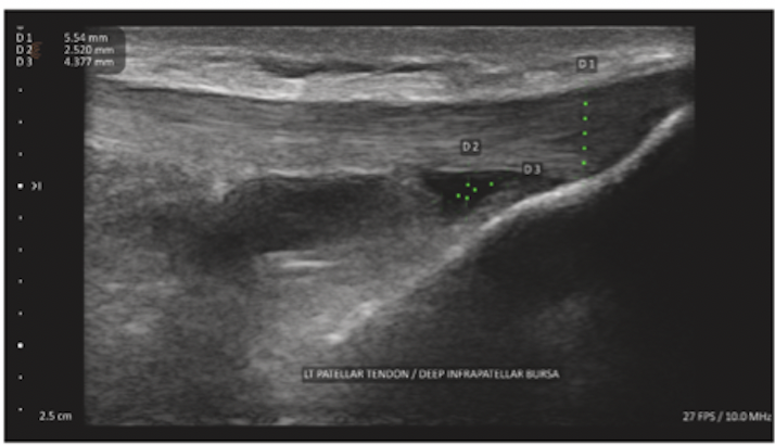
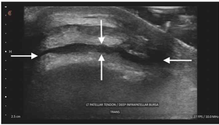
Deep infrapatellar bursal fluid is located posterior to the distal patellar tendon and anterior to the tibia. Recognizing this relationship helps distinguish a bursal collection from tendon disease or joint fluid.
Source: Sachdeva S, Rao VV. Point-of-Care Ultrasound of the Knee: Diagnostic Applications, Procedures, and Best Practices. Journal of Radiology Nursing. 2026;48:101467. Figure 4. https://doi.org/10.1016/j.jradnu.2026.101467
Source: Sachdeva S, Rao VV. Point-of-Care Ultrasound of the Knee: Diagnostic Applications, Procedures, and Best Practices. Journal of Radiology Nursing. 2026;48:101467. Figure 5. https://doi.org/10.1016/j.jradnu.2026.101467
Assess tendon architecture and continuity
Follow the quadriceps tendon to the superior patellar pole and the patellar tendon from the inferior patellar pole to the tibial tuberosity.
Document:
Whether changes are insertional or within the tendon substance.
Tendon thickness and fibrillar architecture.
Focal defects and the extent of fiber disruption.
Whether continuity is preserved.
Associated fluid and Doppler findings, when assessed.
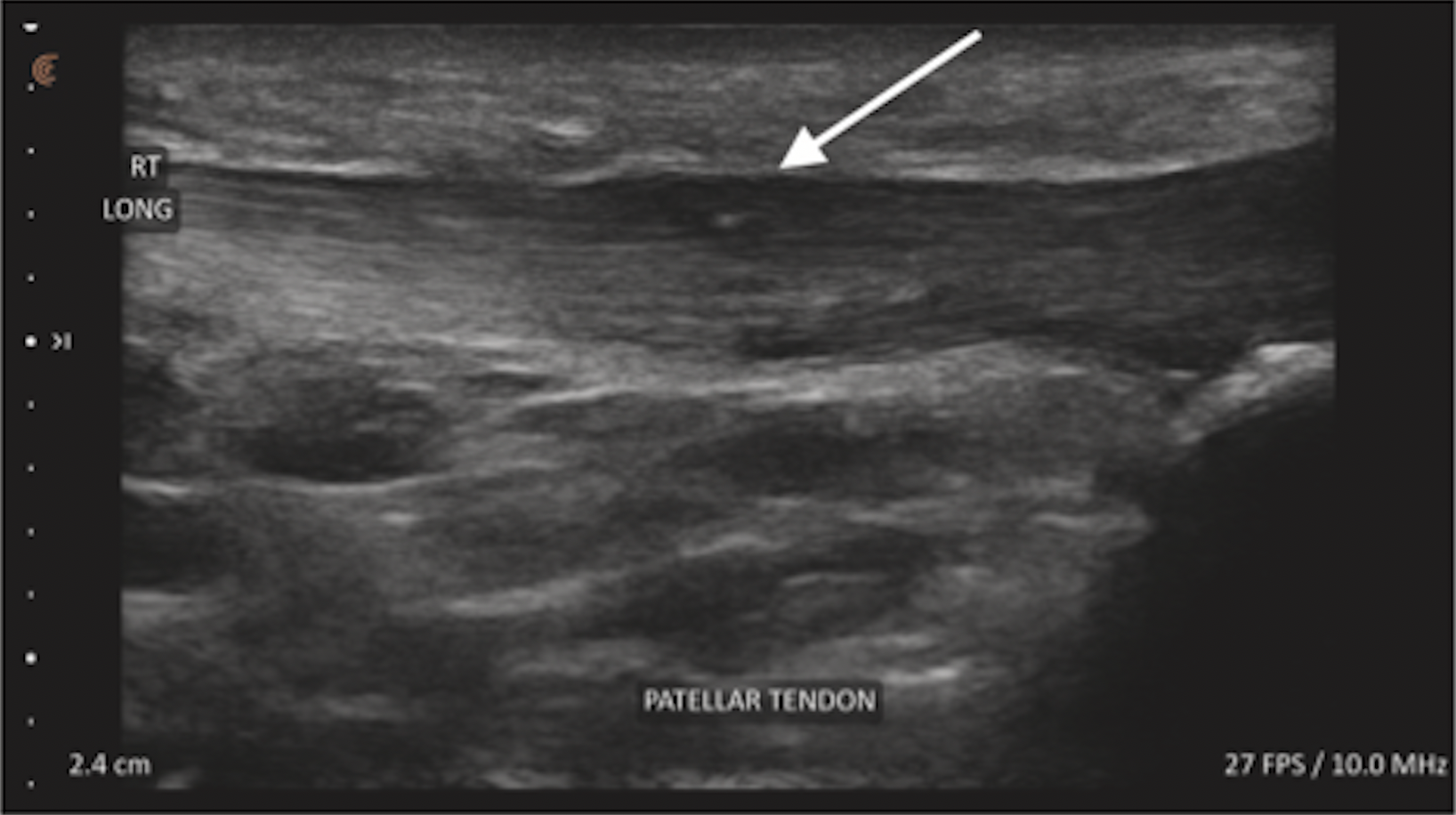
Thickening, hypoechogenicity, and altered fibrillar architecture can accompany tendinopathy. These findings require correlation with symptom location, loading history, and examination findings.
A tear assessment should focus on demonstrable fiber disruption, its extent, and residual intact tissue. Avoid diagnosing a tear from hypoechogenicity alone.
When complete rupture is suspected, integrate imaging with the clinical assessment of the extensor mechanism and arrange appropriate specialist evaluation.
Source: Sachdeva S, Rao VV. Point-of-Care Ultrasound of the Knee: Diagnostic Applications, Procedures, and Best Practices. Journal of Radiology Nursing. 2026;48:101467. Figure 7. https://doi.org/10.1016/j.jradnu.2026.101467
Interpret meniscal and collateral ligament findings within the acoustic window
Ultrasound can assess the accessible portions of the collateral ligaments and peripheral menisci. The medial collateral ligament and underlying peripheral medial meniscus provide useful anatomical landmarks at the medial joint line.However, visualizing peripheral meniscal tissue is not equivalent to evaluating the entire meniscus.
A diagnostic study by Elshimy and colleagues reported encouraging results for meniscal and collateral ligament assessment.[2] Its findings should be interpreted within the study’s population, operator expertise, and reference standard. They do not establish that a negative ultrasound excludes all meniscal pathology.
Persistent mechanical symptoms, suspected deep intra-articular injury, or discordance between the examination and ultrasound may justify MRI or specialist assessment.
In reporting, distinguish a demonstrated abnormality from a structure that was incompletely visualized.
Recognize a Baker’s cyst through its anatomical relationship
A typical Baker’s cyst arises in the gastrocnemius–semimembranosus bursa. Examine the posteromedial knee and identify the relationship between the collection, semimembranosus tendon, and medial gastrocnemius head.
Document its extent, internal contents, and any visible communicating neck. Posterior acoustic enhancement supports a fluid-containing structure but is not specific to a Baker’s cyst.
A meta-analysis of 13 studies involving 1,011 participants found high diagnostic accuracy for ultrasound in identifying whether a Baker’s cyst was present.[3] This does not establish that every posterior knee collection is a Baker’s cyst or that an identified cyst explains all symptoms.
A Baker’s cyst can coexist with other pathology. New calf swelling or clinical concern for deep vein thrombosis requires appropriate vascular assessment.
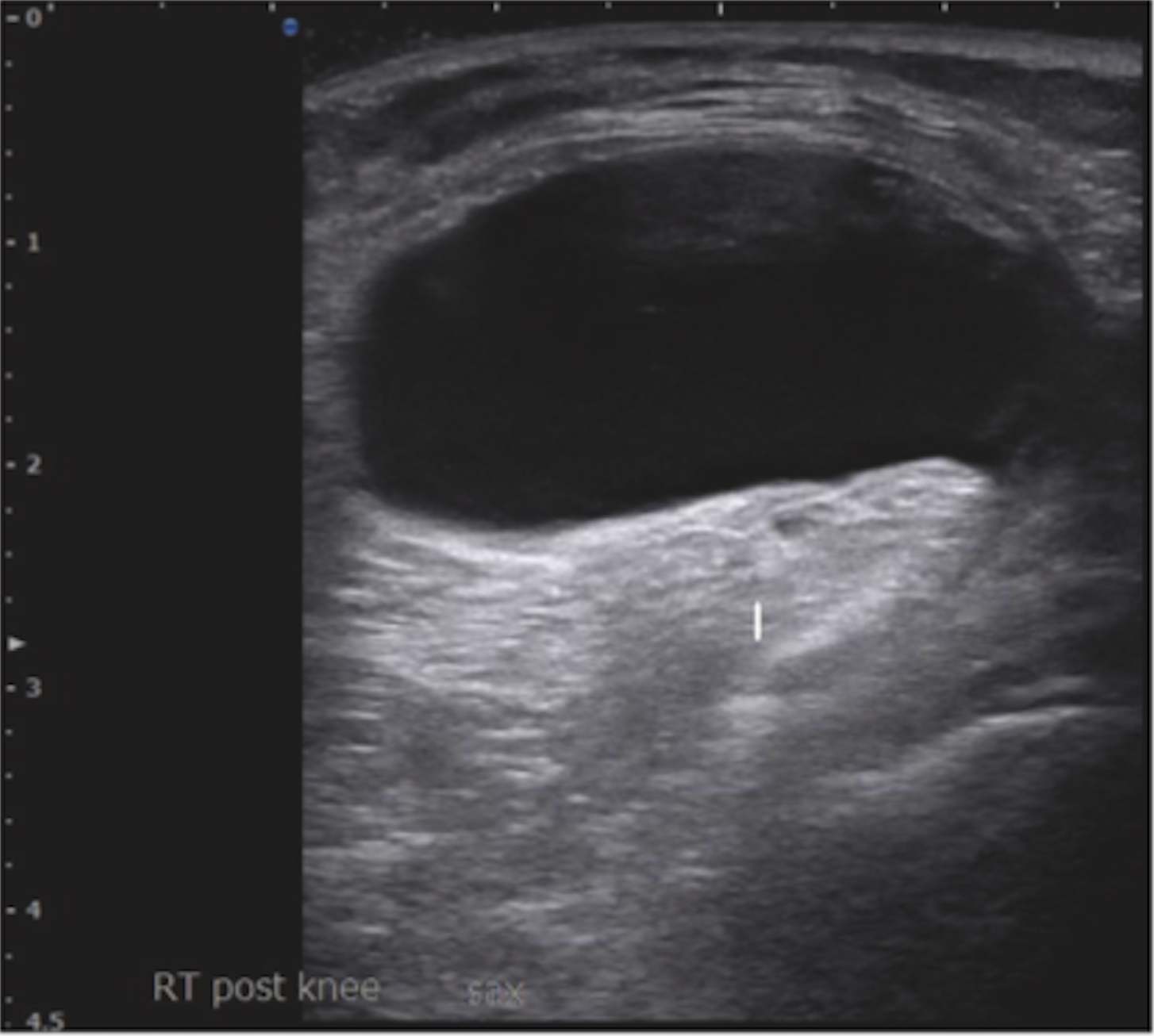
Source: Sachdeva S, Rao VV. Point-of-Care Ultrasound of the Knee: Diagnostic Applications, Procedures, and Best Practices. Journal of Radiology Nursing. 2026;48:101467. Figure 9. https://doi.org/10.1016/j.jradnu.2026.101467
Report the finding and its limitations
An actionable report should state:
Location: side, structure, and anatomical level.
Appearance: architecture, echogenicity, fluid, and vascularity where assessed.
Extent: dimensions and structural continuity when relevant.
Clinical correlation: whether the finding corresponds to the symptomatic region.
Limitations: structures or pathology the examination could not adequately assess.
For example, describing “fluid distension of the deep infrapatellar bursa” communicates more useful anatomical information than “fluid around the knee.”
Similarly, a report should not imply that internal derangement has been excluded when the examination addressed only superficial structures.
Apply the review with appropriate clinical judgment
The source article also discusses ultrasound-guided procedures and emerging AI applications. Procedural visualization requires separate competencies in needle tracking, anatomical safety, and infection prevention.
AI-generated labels or overlays may assist orientation, but an illustrative image does not demonstrate validated diagnostic performance. The clinician remains responsible for confirming the anatomy and interpreting the underlying ultrasound image.
The practical value of knee ultrasound comes from connecting a specific clinical question with a technically sound examination and an appropriately qualified interpretation.
Advance your MSK ultrasound practice
Pura Health’s Lower Extremity Soft Tissue & Ligament Ultrasound course connects regional anatomy, scanning technique, and clinical interpretation for sports medicine clinicians.
Explore ultrasound education with Pura Health
References
Sachdeva S, Rao VV. Point-of-Care Ultrasound of the Knee: Diagnostic Applications, Procedures, and Best Practices. Journal of Radiology Nursing. 2026;48:101467. https://doi.org/10.1016/j.jradnu.2026.101467
Elshimy A, Osman AM, Awad MES, Abdel Aziz MM. Diagnostic accuracy of point-of-care knee ultrasound for evaluation of meniscus and collateral ligaments pathology in comparison with MRI. Acta Radiologica. 2023;64(7):2283–2292. https://doi.org/10.1177/02841851211058280
Liu K, Li X, Weng Q, Lei G, Jiang T. Diagnostic accuracy of ultrasound for the assessment of Baker’s cysts: a meta-analysis. Journal of Orthopaedic Surgery and Research. 2022;17:535. https://doi.org/10.1186/s13018-022-03430-9