
Introduction
Diagnostic ultrasound is traditionally associated with evaluation of muscles, tendons, ligaments, nerves, and superficial joints.
However, growing evidence suggests that ultrasound may also provide valuable information regarding fracture healing and bone remodeling.
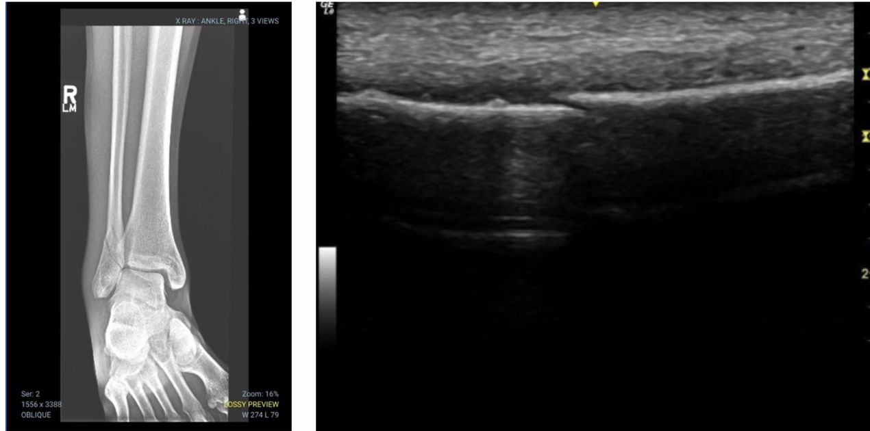
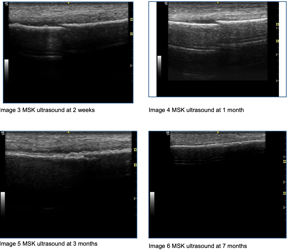
A recent case report followed a distal fibular fracture using serial ultrasound examinations at:
2 weeks
1 month
3 months
7 months
demonstrating progressive healing and callus formation over time.
Why Fracture Follow-Up Matters
Patients often return to activity before complete healing has occurred.
This is particularly relevant for:
Fibular fractures
Fifth metatarsal fractures
Stress injuries
Athletes returning to sport
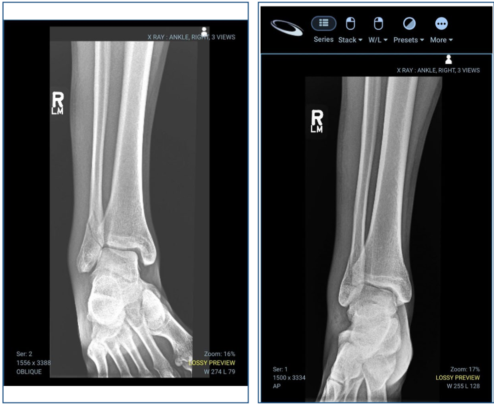
Traditionally, monitoring relies on radiography.
However:
repeated radiation exposure
cost
accessibility
remain limitations.
What Can Ultrasound See?
Normal cortical bone appears as:
✔ bright hyperechoic cortical line✔ posterior acoustic shadow
When a fracture occurs:
✔ cortical disruption develops✔ irregularity becomes visible✔ fracture gap may be identified
As healing progresses:
✔ soft callus develops✔ mineralization increases echogenicity✔ cortical continuity gradually returns
This allows ultrasound to visualize the biological healing process in real time.
Understanding the Stages of Healing
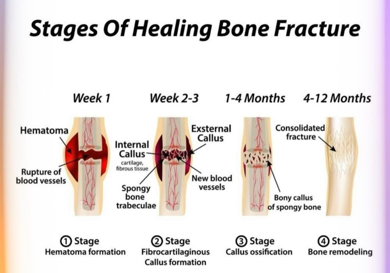
1. Inflammatory Phase
hematoma formation
inflammatory signaling
cellular recruitment
2. Reparative Phase
soft callus formation
fibrocartilage development
early ossification
3. Remodeling Phase
woven bone replaced
cortical architecture restored
mechanical strength improves
Ultrasound findings evolve alongside these biological stages.
Potential Advantages of Ultrasound
Ultrasound offers several practical advantages:
✔ no ionizing radiation✔ portable✔ relatively inexpensive✔ repeatable serial assessments✔ real-time imaging
Previous studies have even suggested that ultrasound may identify callus formation before radiographic evidence becomes apparent.
Clinical Implications for Sports Medicine
For sports medicine clinicians, diagnostic ultrasound may provide an additional tool to:
monitor healing progression
guide loading decisions
assess cortical remodeling
improve patient education
support return-to-activity discussions
The future role of ultrasound in fracture management may extend beyond diagnosis into longitudinal monitoring.
Conclusion
Diagnostic ultrasound has become an indispensable tool for evaluating soft tissue injuries.
Emerging evidence suggests it may also provide meaningful information regarding fracture healing and bone remodeling.
While radiography remains the standard for fracture diagnosis and follow-up, ultrasound offers a radiation-free, accessible, and dynamic adjunct that may help clinicians monitor healing progression over time.
As ultrasound technology and research continue to evolve, its role in musculoskeletal medicine may extend well beyond tendons and ligaments.