
Carlos Jimenez, DPT, RMSK, CPSS, PhD(c) | Founder, Pura Health
A useful ultrasound assessment of a calf injury should describe the involved muscle, the injured myoconnective structure, and the extent of architectural disruption. These findings help clinicians establish a more precise diagnosis and identify questions that require further investigation.
The 2026 review by Pedret and colleagues emphasizes the value of this anatomical approach while acknowledging an important limitation: increasingly detailed imaging does not yet provide reliable, individualized return-to-sport predictions.[1]
For clinicians performing musculoskeletal ultrasound, the challenge is to translate structural findings into appropriate clinical decisions.
Identify the injured structure
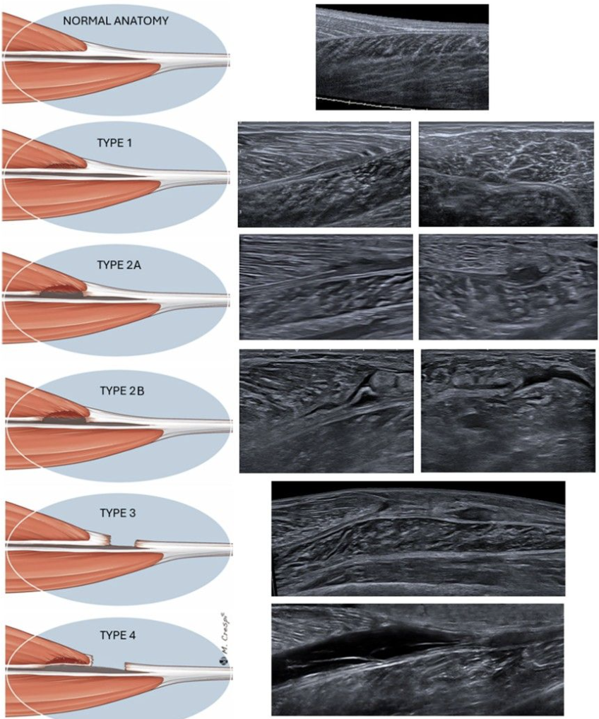
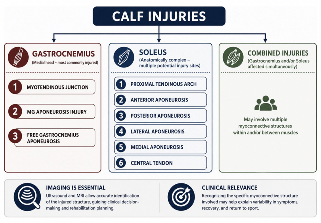
The gastrocnemius–soleus complex contains several potential injury sites. A report describing only a “medial calf tear” may overlook clinically relevant distinctions.
For the medial gastrocnemius, distinguish involvement of:
Muscle fibers and the myotendinous junction.
The gastrocnemius aponeurosis.
The free aponeurosis distal to the muscle fibers.
Combined muscular and aponeurotic injury.
The soleus has a more complex internal architecture, including a central tendon, several aponeurotic components, and a proximal tendinous arch. Its deeper location and anatomical variability can make ultrasound assessment challenging.
Precise localization improves communication between the imaging clinician and rehabilitation team. However, an anatomical label alone does not establish injury severity, functional capacity, or a recovery timeline.
Build the examination around anatomy and symptoms
Start with the injury mechanism, symptom distribution, focal tenderness, and response to loading. Use these findings to guide the examination while assessing the surrounding anatomy.
A practical ultrasound examination should:
Establish orientation with a regional survey.
Follow the relevant muscle and connective tissue structures proximally and distally.
Examine abnormalities in both longitudinal and transverse planes.
Assess adjacent structures and the gastrocnemius–soleus interface.
Use gentle dynamic assessment when appropriate and tolerated.
Optimize frequency, depth, and focal position for the structure being examined. A setting suitable for superficial gastrocnemius may provide inadequate visualization of the deeper soleus.
Before interpreting a hypoechoic region as injury, adjust the insonation angle. Anisotropy can alter the appearance of tendinous and aponeurotic structures. Confirm a suspected abnormality across planes and determine whether there is genuine architectural disruption.
Contralateral comparison can help clarify anatomy, but should complement a technically adequate examination.
Describe the sonographic findings precisely
The examination should establish what is disrupted, where the disruption occurs, and how confidently its extent can be assessed.
Document:
Reporting domain | Information to include |
Localization | Muscle, side, proximal–distal location, and anatomical landmark |
Structural involvement | Muscle fibers, myotendinous junction, aponeurosis, or tendon |
Architecture | Preserved or disrupted fibers; connective tissue continuity |
Extent | Dimensions in specified planes and visible retraction, if present |
Associated findings | Fluid collection and its anatomical compartment |
Examination limitations | Inadequately visualized structures or uncertain injury extent |
Fluid between the medial gastrocnemius and soleus should prompt careful assessment of the adjacent structures. Its presence alone does not identify the full injury pattern.
When using an imaging classification, state which system you are applying. Classification terminology should accompany a clear anatomical description.
Recognize the limits of ultrasound in soleus injury
A technically satisfactory superficial examination does not exclude a deeper soleus injury.
Consider whether the examination adequately visualized the symptomatic region, internal connective tissue structures, and suspected proximal extent. Early injuries may also have subtle findings.
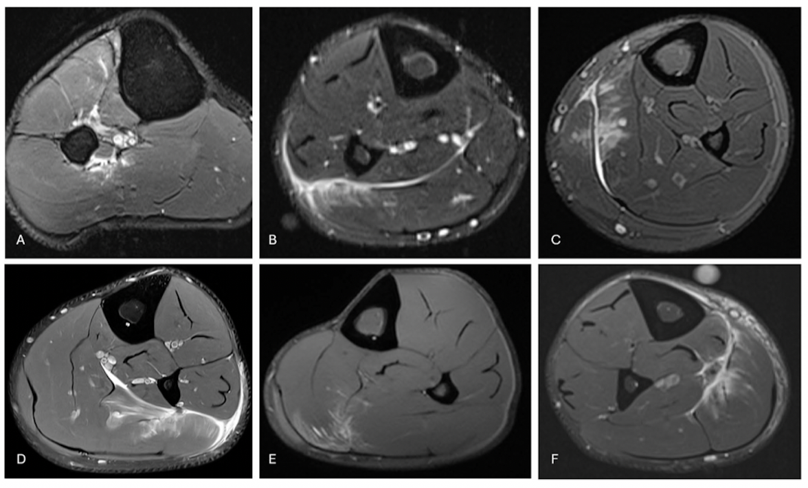
MRI may be useful when:
Symptoms suggest deep soleus involvement that ultrasound cannot characterize.
Clinical findings and ultrasound findings are discordant.
The suspected injury extends beyond the accessible acoustic window.
Clarifying a complex injury would change management.
Additional imaging should answer a specific clinical question. Greater anatomical detail does not au
tomatically provide greater prognostic certainty.
Keep the differential diagnosis open
Posterior calf pain includes muscular, tendinous, and nonmusculoskeletal causes. Relevant alternatives include Achilles tendon injury, plantaris injury, a ruptured popliteal cyst, and deep vein thrombosis.[2]
A focused musculoskeletal scan does not substitute for an appropriate venous assessment when DVT is suspected. Severe or atypical symptoms should also prompt consideration of conditions requiring urgent evaluation, including acute compartment syndrome.
Identifying a muscle injury should not prematurely close the diagnostic process when the clinical presentation remains unexplained.
Connect imaging to rehabilitation decisions
Anatomical findings can help guide the emphasis and monitoring of rehabilitation.
Assess plantar-flexor capacity with both the knee extended and flexed. These positions alter muscular contributions, but neither perfectly isolates a muscle nor identifies an injury subtype.
Progressive rehabilitation should address:
Force production and repeated-force capacity.
Loading through relevant muscle–tendon lengths.
Rapid force development and elastic loading.
Running volume, speed, and acceleration.
Deceleration, direction changes, and repeated sport-specific efforts.
Aponeurotic or tendinous involvement may warrant closer monitoring as mechanical demands increase. It does not establish a universal period of unloading or a predetermined return date.
For serial ultrasound, use reproducible positioning, imaging planes, and settings where possible. Interpret apparent structural changes alongside symptoms and function. Residual imaging abnormalities may persist despite clinical improvement.
Separate structural assessment from return-to-sport clearance
Ultrasound contributes information about tissue structure. Return-to-sport decisions also require evidence that the athlete can tolerate the intended demands.
Integrate examination findings, strength and endurance, elastic loading, running exposure, sport-specific performance, and athlete confidence. Expert clinical practice supports individualized progression and repeated assessment of responses to loading.[3]
The 2026 review identifies no validated calf-specific rehabilitation or return-to-sport criteria.[1] A favorable scan cannot independently establish readiness, and persistent imaging changes do not automatically prohibit progression.
The value of an advanced ultrasound examination lies in precise anatomical characterization, recognition of uncertainty, and meaningful integration with clinical assessment.
Develop your clinical ultrasound skills
Pura Health’s Lower Extremity Soft Tissue & Ligament Ultrasound course connects regional anatomy, practical scanning, and clinical interpretation for sports medicine professionals.
Explore Pura Health ultrasound education
References
Pedret C, et al. Calf Injuries: An Anatomy-Based Approach to Diagnosis, Management and Return to Sport. Current Reviews in Musculoskeletal Medicine. 2026. Read the review.
Bright JM, Fields KB, Draper R. Ultrasound Diagnosis of Calf Injuries. Sports Health. 2017;9(4):352–355. Read the article.
Green B, et al. The Assessment, Management and Prevention of Calf Muscle Strain Injuries: A Qualitative Study of the Practices and Perspectives of 20 Expert Sports Clinicians. Sports Medicine - Open. 2022;8:10. Read the study.