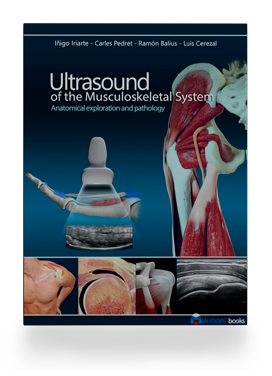
RECOMMENDED BOOK
Ultrasound of the Musculoskeletal System
Iriarte, Pedret, Balius & Cerezal
View the book
YOUR COURSE
PURA HEALTH
Learn. Scan. Apply.
Choose a section to start learning or pick up where you left off.
WELCOME BACK
Use the Pura Health account you enrolled with to open your course sections.
Checking your course access…
This account doesn’t have an active Lower Extremity enrollment. If your team provided a code, apply it when enrolling.
Use my training codeGet access helpWELCOME TO YOUR COURSE
Instructor: Carlos Jimenez, DPT, RMSK, CPSS. Disclosure: Carlos Jimenez reports nothing to disclose.
Open Ultrasound Fundamentals, choose the first video, and press play. You can pause whenever you need—your viewing progress saves to your account.
Open your first lesson →YOUR NEXT STEP
Choose a section below while your saved progress loads.
After completing all eight sections, submit your clinical reflection for review, then complete the course evaluation.
Reflection, evaluation & certificateTHE LEARNING PATH
Start with Fundamentals or choose a region to review.
Probe orientation, image optimization, and the settings behind a clear musculoskeletal scan.
Rectus femoris zones, central tendon patterns, and their clinical relevance.
The MCL, patellar tendon, joint recesses, and fat pads in the athletic knee.
Key tendon landmarks and strain-prone zones in the proximal adductors.
Iliopsoas, sartorius, and anterior joint landmarks on ultrasound.
Proximal hamstring tendons and common strain locations.
Ankle ligaments and tendons, plus the gastrocnemius–soleus complex.
Muscle–tendon behavior during movement and loading, alongside static imaging.
KEEP THESE CLOSE
RECOMMENDED BOOK
Iriarte, Pedret, Balius & Cerezal
View the bookINSIDE THE COURSE SECTIONS
Open a section to find its existing learning materials, including video walkthroughs and supporting resources.
BRING IT INTO PRACTICE
Track your submissions, scores, and instructor feedback.
Prepare your scan images, video clips, and report for review.